
Primary Ciliary Dyskinesia: When the Body’s Tiny Cilia Don’t Work as They Should
Imagine hundreds of microscopic hairs lining your airways, all beating together like synchronized swimmers. Their job is to move mucus, along with trapped bacteria, particles, and debris, upward toward the throat, where it can be swallowed or coughed out.
These tiny, hairlike structures are called cilia.
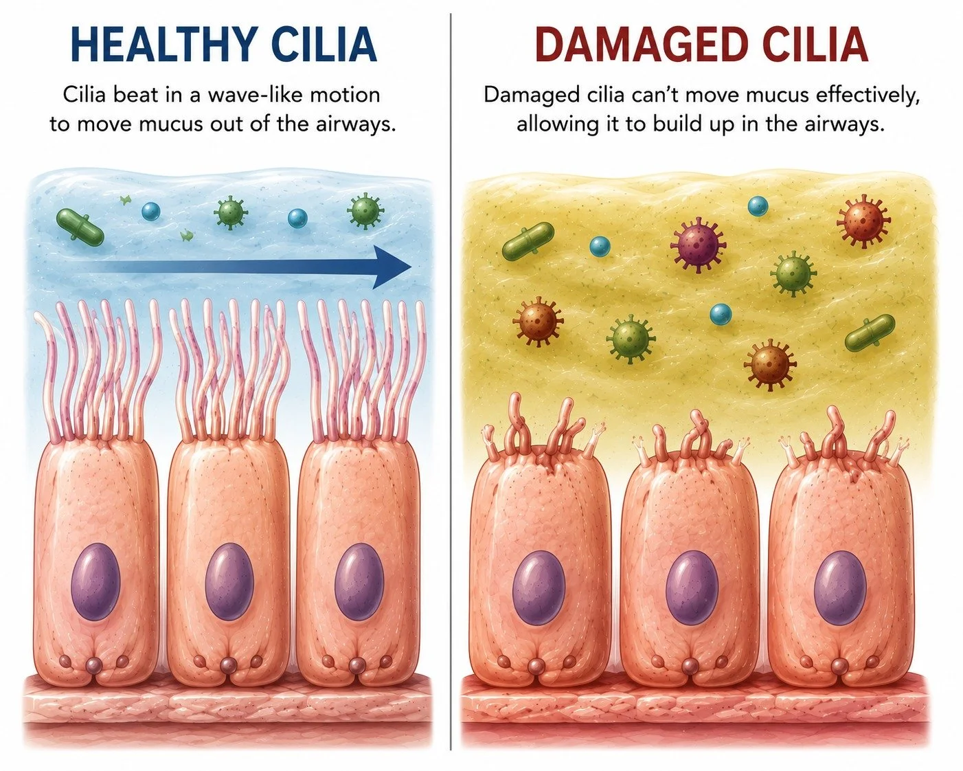
In primary ciliary dyskinesia, or PCD, inherited genetic changes cause the cilia to be missing, structurally abnormal, poorly coordinated, or unable to move effectively. I think of synchronized swimmers who are not doing a very good job of staying in sync!
Without this natural sweeping action, mucus and bacteria can remain in the airways. Over time, this can contribute to repeated infections, chronic inflammation, and bronchiectasis.
Bringing the Global PCD Community Together
PCD was recently the focus of the inaugural Global PCD Conference in Montreal, Canada. Clinicians, researchers, people living with PCD, patient advocacy organizations, and others interested in advancing our understanding of this rare disease came together to learn, share their knowledge, and encourage greater collaboration.
Among the international experts participating were Professor James D. Chalmers of the University of Oxford and Professor Amelia Shoemark of the University of Dundee in Scotland. Professor Shoemark has been instrumental in identifying several of the genes now known to cause PCD.
Also attending was Judy Schloss, RRT, Clinical and Medical Director at Monaghan Medical Corporation, the maker of the Aerobika® OPEP device and the AeroEclipse® breath-actuated nebulizer.
Judy shared something with me that I found especially interesting. Researchers are examining data from a subgroup of 117 participants living with both PCD and bronchiectasis, drawn from the more than 1,700 participants in the Phase 3 ASPEN study of brensocatib.
It will be interesting to learn what impact, if any, this new medication had on people living with these overlapping conditions.
What Is Primary Ciliary Dyskinesia?
PCD is a rare, inherited disorder affecting motile cilia. These cilia are found throughout the respiratory tract, as well as in the middle ear, reproductive system, and other areas of the body.
In healthy airways, hundreds of cilia on each ciliated cell beat together in coordinated waves. This movement is part of our natural defense system, continually helping to move mucus and anything trapped within it up and out of our lungs.
In PCD, the cilia cannot perform this job effectively. Mucus may remain in the airways, creating an environment in which bacteria can become trapped, and infections can develop.
This is why many people with PCD experience a daily wet cough, chronic sinus and ear problems, repeated respiratory infections, and, over time, bronchiectasis.
Signs and Symptoms Often Begin Early
PCD is present from birth, although some people are not diagnosed until adulthood.

Common signs and symptoms may include:
Respiratory distress in an otherwise full-term newborn
A daily wet or productive cough beginning in infancy
Year-round nasal congestion and chronic sinusitis
Recurrent middle ear infections
Possible hearing loss
Repeated chest infections
Bronchiectasis, which may develop over time
Reduced fertility in some women
Reduced fertility or infertility in many men
Differences in the placement of internal organs in some people
Not everyone with PCD will experience every one of these problems, and the severity of the disease can vary considerably.
PCD and the Placement of Our Organs
Cilia also play an important role very early in embryonic development. Specialized cilia help establish the left-right orientation of the developing organs.
When these cilia do not function properly, the usual pattern may not be established. As a result, approximately 40% to 50% of people with PCD have situs inversus, in which the major internal organs are arranged as a mirror image of their usual positions. For example, the heart may be on the right side of the chest rather than the left.
When someone has PCD, situs inversus, and the characteristic respiratory features of chronic sinus disease and bronchiectasis, the combination has historically been called Kartagener syndrome.
Kartagener syndrome is considered a clinical subset of PCD, not a separate cause of ciliary dysfunction.
I have worked with people whose Kartagener syndrome was not discovered until adulthood, sometimes when an X-ray unexpectedly revealed that the heart and other organs were positioned on the opposite side.
It is important to note that most people with PCD do not have the classic combination known as Kartagener syndrome. Having organs in their usual positions certainly does not rule out PCD.
How Is PCD Diagnosed?
PCD can be challenging to diagnose because there is no single test that identifies every case.
An evaluation generally begins with a detailed clinical history, paying particular attention to symptoms present at birth or in early childhood. A clinician may also review imaging, lung function results, previous respiratory cultures, and a person’s history of sinus and ear disease.
Diagnostic testing may include:
Genetic testing, often using a multigene PCD panel
Nasal nitric oxide measurement
Transmission electron microscopy to examine the structure of the cilia
High-speed video microscopy to evaluate ciliary movement
Immunofluorescence testing to look for missing or misplaced ciliary proteins
Genetic testing is now a central part of the diagnostic evaluation. A diagnosis may be confirmed when disease-causing variants are identified in a gene known to cause PCD and the findings fit the person’s clinical history.
However, a negative or inconclusive genetic test does not necessarily exclude PCD. Researchers are continuing to identify additional genes and disease-causing variants.
Nasal nitric oxide is usually very low in people with PCD and can be a valuable screening tool when it is performed by trained personnel using standardized procedures. It is not diagnostic by itself, and some people with PCD may have nasal nitric oxide levels that are within the normal range.
To have my own nasal nitric oxide test, I went to a children’s center here in New York City. I was quite surprised when I walked in and saw little tables and chairs and a guitar player performing “Puff, the Magic Dragon.”
It was a memorable experience, but it also illustrated an important point. We often need to go to pediatric or specialty centers because they have the equipment and experience required to perform PCD testing properly.
In some communities, this type of specialized testing may be difficult, if not impossible, to access. Someone may need to travel a considerable distance to reach an experienced center.
Transmission electron microscopy, high-speed video microscopy, and immunofluorescence testing can also provide valuable information, but each has limitations. For example, some people with PCD have ciliary defects that cannot be seen with electron microscopy. Ciliary movement may also be temporarily altered by infection or another acquired condition.
This is why test results need to be considered together rather than in isolation.
Clearly, diagnosing PCD can be complicated. Whenever possible, someone with suspected PCD should be evaluated at a center with specific expertise in diagnosing and managing the disease.
How Is PCD Managed?
Unfortunately, there is currently no cure for the underlying ciliary dysfunction.
Management, which shares many similarities with the management of bronchiectasis, focuses on protecting the lungs, reducing infections, and helping to compensate for the work the cilia are unable to do.
Because the cilia cannot move mucus effectively, airway clearance is especially important. The goal is to loosen, mobilize, and remove mucus before it accumulates and contributes to infection, inflammation, and further airway damage. Devices such as an Aerobika or AeroEclipse XL are often used for this, and keeping them clean and properly disinfected is an important part of using them safely.

A person’s treatment plan may include:
A regular, individualized airway-clearance routine
Exercise and physical activity, when appropriate
Sputum cultures and lung function testing
Prompt identification and treatment of respiratory infections
Monitoring and treatment of sinus and ear disease
Hearing evaluations
Recommended vaccinations
Fertility counseling or support when needed
Treatment should always be individualized with the guidance of clinicians who understand PCD and bronchiectasis.
Why Greater Awareness Matters
PCD is rare, its symptoms can vary, and diagnosis is frequently delayed. Some adults have lived with a chronic wet cough, sinus disease, ear infections, or repeated chest infections since childhood without anyone putting all the pieces together.
We also know that PCD may be an underrecognized underlying cause of bronchiectasis.
This is why determining the underlying cause of bronchiectasis matters. When PCD is identified, it can help guide treatment, monitoring, family counseling, and decisions about whether relatives should also be evaluated.
Conferences such as the Global PCD Conference bring clinicians, researchers, people living with the disease, advocates, and industry representatives together. They encourage collaboration, deepen our understanding of PCD, and help move research toward better diagnostic tools and new treatments.
For those living with PCD and bronchiectasis, that growing attention offers something especially important: hope.
Want to Learn More about Bronchiectasis?
The BE CLEAR® Guide to Bronchiectasis is designed to help you understand and manage your chronic lung condition at your own pace. Whether you’re newly diagnosed or looking to strengthen your daily routine, the guide offers expert-informed content, practical suggestions, and compassionate support every step of the way.
